
The Screening Conversation Is Changing
For decades, mammography has been the cornerstone of breast cancer screening in the United States. It has saved lives — that’s not in dispute. But it has also had well-documented limitations that women and their doctors have navigated with varying degrees of success. Dense breast tissue. Compression discomfort. Overlapping structures that obscure findings. Callback rates that generate anxiety without always generating answers.
The imaging field has been working on better solutions for years. Tomosynthesis — 3D mammography — was a meaningful step forward. MRI offers exceptional soft-tissue detail but comes with cost, access, and contrast injection barriers. Ultrasound is useful as a supplement but not designed as a standalone screening tool.
Now, a technology that’s been gaining serious clinical traction is drawing increased attention from radiologists, breast surgeons, and oncologists across the US: 3D breast CT. It’s not yet universally available, and it’s not positioned as a replacement for every existing modality — but for the right patients and clinical questions, it represents a genuinely significant advance in how we image the breast.
This blog is for women who want to understand what this technology is, how it works, and what it might mean for their own screening or diagnostic journey.
The Core Problem With Conventional Breast Imaging
To appreciate what 3D breast CT offers, it helps to understand the fundamental limitation it addresses.
Conventional mammography is a projection imaging technique. It compresses the breast and captures a two-dimensional image of a three-dimensional structure. The compression serves a purpose — it spreads tissue apart and reduces the thickness the X-ray must penetrate — but it has a significant consequence: structures overlap. A benign cyst positioned in front of or behind a small mass can obscure it entirely. A dense fibroglandular region can hide a lesion in plain sight.
This is the core problem that drove the development of tomosynthesis, which acquires multiple low-dose images from different angles and reconstructs them into quasi-3D slices. It helps — studies have consistently shown improved cancer detection rates and reduced recall rates compared to 2D mammography. But it’s still a limited-angle acquisition, and the reconstruction artifacts it produces can be clinically significant.
True volumetric imaging — acquiring a genuine three-dimensional dataset of the entire breast — is a fundamentally different approach. And that’s what a dedicated breast CT system delivers.
How Dedicated Breast CT Actually Works
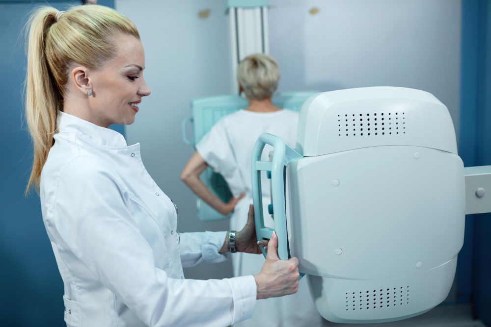
A dedicated breast CT system is designed specifically for breast imaging, which distinguishes it from whole-body CT scanners adapted for breast use. The patient lies prone on a specialized table with the breast positioned in a pendant configuration through an opening in the table — no compression required. A cone-beam CT acquisition rotates around the breast, acquiring hundreds of projection images that are reconstructed into a true isotropic 3D volume.
The result is a complete volumetric dataset with sub-millimeter resolution in all three dimensions. Radiologists can navigate through the breast in any plane — axial, coronal, sagittal, or oblique — and examine structures without the overlay problem that fundamentally limits planar projection imaging.
Why no compression matters more than you might think
The absence of compression in dedicated breast CT isn’t just about patient comfort, though that matters too. It means the breast is imaged in a more natural configuration, without the tissue distortion that compression creates. Structures are where they actually are. Relationships between lesions and surrounding tissue are preserved. For surgical planning and for patients with implants, this is clinically meaningful.
For women who have avoided or delayed screening because compression is genuinely painful — a more common experience than often acknowledged, particularly for women with dense or sensitive breast tissue — the comfort difference can be the factor that makes consistent screening actually achievable.
What 3D Breast CT Sees Differently
The clinical value of 3D breast CT shows up most clearly in specific imaging scenarios.
Dense breast tissue
Dense breast tissue is one of the most significant challenges in mammographic screening. It’s both a masking factor — dense tissue can hide cancers — and an independent risk factor for breast cancer development. Approximately 40 to 50 percent of women in the US have dense breasts, and many receive supplemental screening recommendations as a result.
Because breast CT acquires a true volumetric dataset rather than a projection image, density doesn’t create the same masking problem. Structures within dense tissue can be visualized in 3D, reducing the probability that a lesion is hidden simply because it’s surrounded by fibroglandular tissue.
Mass characterization
When a mass is detected, understanding its three-dimensional morphology — its shape, margins, internal architecture, and relationship to surrounding structures — is critical for determining its significance. In 2D mammography, you get two projections. In tomosynthesis, you get limited-angle reconstruction. In dedicated breast CT, you get the complete three-dimensional structure of the mass from every angle simultaneously.
This level of detail supports more confident characterization and can reduce the rate of unnecessary biopsies for lesions that are clearly benign on volumetric imaging but ambiguous on planar views.
Calcification assessment
Calcifications — tiny calcium deposits in breast tissue — are one of the most common mammographic findings and one of the most nuanced to interpret. Their morphology, distribution, and clustering pattern all factor into the assessment. Volumetric CT imaging provides the full three-dimensional distribution of calcification clusters, which adds information that 2D projections and even tomosynthesis can’t fully capture.
The Radiation Question
Any conversation about CT imaging appropriately includes a discussion of radiation dose. CT has historically been associated with higher radiation exposure than mammography, and that concern is legitimate in the context of whole-body CT.
Dedicated breast CT systems have been specifically engineered to deliver doses comparable to or in some configurations lower than standard two-view mammography. The technology uses optimized acquisition protocols, specialized detector systems, and targeted reconstruction algorithms designed for the specific geometry of breast imaging — not adapted from body CT.
As with any imaging modality, the dose consideration has to be weighed against the diagnostic benefit for each individual patient and clinical indication. For the right patient and question, a breast ct scan that delivers definitive diagnostic information with acceptable dose is a favorable trade-off compared to multiple inconclusive studies requiring follow-up.
Who Is This Technology Most Relevant For Right Now?
Dedicated breast CT is not currently positioned as a universal screening replacement for mammography. That’s an important clarification. The technology is most relevant in specific clinical contexts, and understanding those contexts helps women have more informed conversations with their care teams.
Problem-solving after inconclusive mammography or tomosynthesis
When a screening study identifies something that requires further evaluation, the diagnostic workup typically involves additional mammographic views, targeted ultrasound, and sometimes MRI. Dedicated breast CT offers an additional problem-solving tool with a different set of strengths — particularly for findings in dense tissue or for masses requiring precise 3D characterization.
Pre-surgical planning
Surgeons benefit from precise three-dimensional information about tumor location, size, and relationship to surrounding structures. Dedicated breast CT provides that information in a format that is often more directly applicable to surgical planning than 2D projections.
Patients for whom MRI is contraindicated or impractical
Breast MRI is a powerful tool, but it’s not available or appropriate for every patient. Women with pacemakers or certain implants, women who experience significant claustrophobia, or women in healthcare systems where MRI access is constrained may benefit from breast CT as an alternative high-detail imaging option.
High-risk screening programs
For women at elevated lifetime risk — due to genetic factors, family history, or prior biopsy findings — supplemental screening beyond standard mammography is often recommended. As the clinical evidence base for dedicated breast CT develops, it is increasingly being explored as a supplemental screening option in this population.
The State of the Technology in the US
Dedicated breast CT systems are FDA-cleared and available at an expanding number of breast imaging centers, academic medical centers, and comprehensive cancer programs across the United States. Availability is growing but not yet universal, and insurance coverage varies by indication and payer.
If you’re interested in whether breast CT might be relevant to your situation, the conversation starts with your primary care physician, gynecologist, or breast imaging specialist — particularly if you have dense breasts, a history of inconclusive mammographic findings, or elevated breast cancer risk.
Take an Active Role in Your Breast Health
Breast imaging technology is advancing, and you deserve to understand your options — not just accept whatever default protocol you’re offered. Ask questions. Understand your breast density. Know your risk factors. And when a new imaging option might offer clearer answers than what you’ve had before, advocate for that conversation.
Talk to your doctor about whether 3D breast CT is right for you. Ask whether it’s available at your imaging center, whether your clinical situation might benefit from volumetric breast imaging, and what the evidence says for your specific risk profile. Informed patients get better care — and better care starts with the right questions.


