
The landscape of skin health monitoring has evolved significantly over the past decade, placing a higher emphasis on non-invasive diagnostic tools that allow for a deeper look at the structure of our skin. For individuals concerned about changes in their skin markings, Dermoscopy Mole Evaluation in Abu Dhabi offers a sophisticated approach to analyzing pigmented lesions. This technology serves as a bridge between a simple visual examination and more invasive procedures, providing a high-resolution view that is not possible with the naked eye alone.
-
Technological advancements in dermatology have enhanced the ability to monitor skin health non-invasively.
-
The primary goal is to provide a detailed analysis of pigmented lesions.
-
This method acts as a critical intermediate step before considering further diagnostic steps.
The Science Behind Dermoscopy
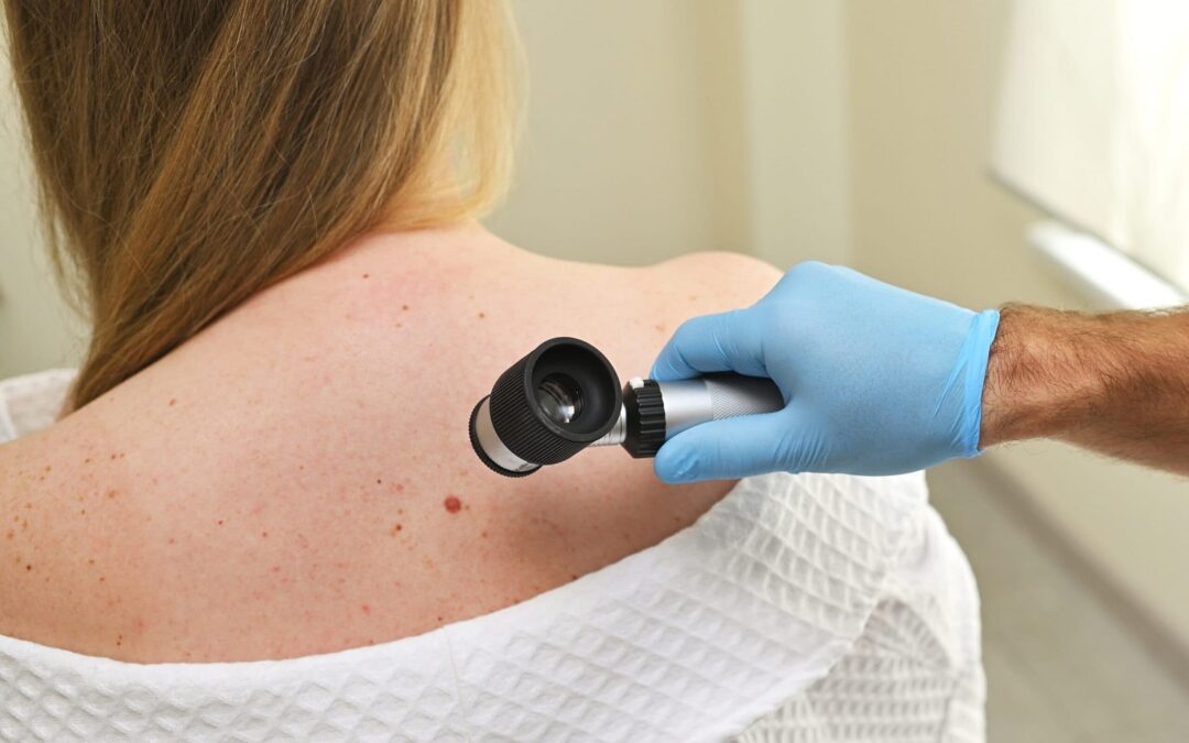
At its core, a dermatoscope is a specialized magnifying device combined with a high-intensity lighting system, often using cross-polarized light to eliminate surface glare. When a professional places this device against the skin, it renders the epidermis transparent, allowing them to visualize subsurface structures, pigment patterns, and vascular arrangements. This process reveals architectural details—such as the distribution of melanin and the integrity of the dermal-epidermal junction—that are otherwise invisible during a standard physical exam.
-
The dermatoscope uses magnification and polarized light to see past the skin’s outer layer.
-
It allows for the examination of vascular and structural patterns of moles.
-
By making the surface transparent, it highlights specific diagnostic criteria like pigment network and streaks.
Why Dermoscopy is Considered Effective
The efficacy of this diagnostic tool lies in its ability to significantly increase the accuracy of identifying suspicious lesions. By utilizing standardized classification systems, such as the pattern analysis method or the ABCD criteria (Asymmetry, Border, Color, Differential structures), observers can systematically categorize moles. This standardized approach reduces subjectivity and provides a common language for tracking changes over time. When combined with digital dermoscopy—where images are stored and compared across multiple visits—the effectiveness of early detection increases dramatically.
-
It moves diagnosis from subjective observation to an objective, criteria-based system.
-
Standardized methods like ABCD help in identifying abnormal features accurately.
-
Digital tracking over time creates a baseline for long-term skin health management.
The Process of Mole Evaluation
When undergoing a mole evaluation, the process is streamlined and comfortable. The skin is usually prepared with a small amount of oil or gel to enhance light transmission, though some modern devices are designed to work without contact. The evaluator systematically checks the lesion, noting its symmetry, border regularity, color distribution, and the presence of any atypical structures. This data is often documented to monitor any evolution in the mole’s appearance during subsequent appointments, which is a key component of preventative skin health.
-
The exam is non-invasive and generally quick to perform.
-
Optical clarity is improved with fluids to minimize reflection.
-
Systematic documentation ensures that future growth or change can be monitored precisely.
Enhancing Early Detection Strategies
The real strength of this evaluation method is its role in proactive health management. Rather than waiting for a lesion to change significantly, current strategies emphasize “total body skin examination” followed by targeted dermoscopic evaluation of specific lesions of interest. By creating a digital map of the skin, the diagnostic team can identify “ugly duckling” lesions—moles that look fundamentally different from the rest of the patient’s baseline. This comparative analysis is highly effective in isolating lesions that require further attention.
-
Proactive scanning is more effective than reactive observation.
-
Digital mapping helps identify outliers or “ugly duckling” moles.
-
Baseline comparisons are the gold standard for tracking skin changes.
Integrating Dermoscopy into Routine Care
Incorporating these evaluations into regular health check-ups is a vital practice for those with a high density of moles or a personal history of skin changes. The objective nature of the examination provides peace of mind through documented evidence. Instead of relying on memory, patients have a visual record that provides a concrete history of their skin’s status. This level of detail allows for informed decision-making regarding skin health and emphasizes the importance of routine surveillance in maintaining long-term well-being.
-
Routine check-ups are essential for individuals with multiple moles.
-
Visual documentation removes the burden of reliance on memory.
-
It fosters a proactive environment for ongoing health maintenance.
The Future of Mole Imaging
As technology progresses, the field is moving toward computer-aided diagnosis (CAD) and artificial intelligence integration. These systems analyze dermoscopic images against thousands of reference cases to provide quantitative feedback. While these tools do not replace professional judgment, they act as a “second set of eyes,” ensuring that subtle patterns are not overlooked. The integration of high-resolution imaging and AI represents the next frontier in improving the diagnostic accuracy of mole evaluations globally.
-
AI and computer-aided diagnostics are becoming standard support tools.
-
Algorithms assist in identifying minute details that might escape human detection.
-
The future of dermatology relies on the synergy between human expertise and machine precision.
Frequently Asked Questions
Is dermoscopy a painful procedure?
No, the procedure is entirely non-invasive and painless. It involves the use of a handheld device placed near or gently against the skin to magnify and analyze the area, requiring no needles or surgical tools.
How often should I have my moles evaluated?
The frequency of evaluations depends on your personal skin history, the number of moles you have, and any changes you may have noticed. It is generally recommended to discuss a customized surveillance schedule with a professional based on your specific profile.
Can dermoscopy diagnose all skin concerns?
While it is an exceptionally powerful tool for evaluating pigmented lesions and identifying structural abnormalities, it is part of a broader diagnostic process. It provides critical visual data that assists professionals in determining the next steps for your skin health.
What should I look for in my own moles between visits?
You should monitor for the “ABCDE” signs: Asymmetry (one half doesn’t match the other), Border (irregular or blurred edges), Color (varied shades), Diameter (larger than a pencil eraser), and Evolving (changing in size, shape, or texture). If you notice any of these, schedule a professional evaluation to discuss your findings.


